Having completed extensive articles on the issues of Pet Geriatrics and Euthanasia, we will commence today with considerations as they relate to specific organs and the diseases associated therewith. We have chosen the eye as the first organ with which to deal.
General considerations
We will deal superficially with the structure of the eye and its accessories. Right at the beginning, we must understand a few things relating to normalcy, so that we can know when there is an abnormal condition.
The eye itself is quite a fascinating piece of equipment. The eyeball, which is lodged in the socket of the skull and held in place by special structures (see below), is divided by the lens and its ligaments into two chambers that contain fluids which ensure the turgor of the ball. Around the eyeball is a quantity of fat which cushions the eye.
 The eye is protected by two eyelids and, in the case of dogs, cats, cows, horses and many other species, there is a “third eyelid” called the nictitating membrane. When a cat awakens after sleep, one can see this “third eyelid” retracting across the eyeball. So many pet owners panic when they see this piece of tissue coming across the eyeball. They think it is an ailment, when in fact it is quite normal.
These eyelids are made up of several layers of tissue. For example, there is the muscular tissue which allows the opening and shutting of the eyelids. Another layer has glands on the free edge of the lid. These can develop into painful swollen cysts. Yet another layer consists of a special membrane called the conjunctiva which covers the surface of the eyeball. This surface often becomes inflamed, and the condition is then known as conjunctivitis, about which we will write on a later date.
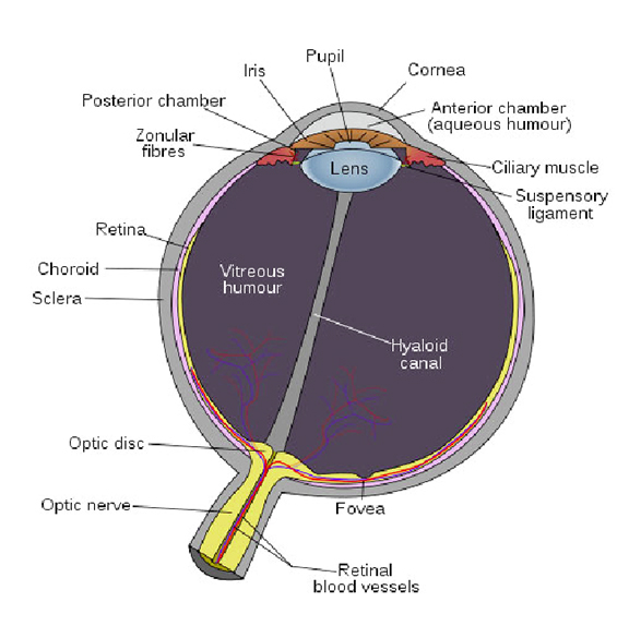
Below the thin conjunctival membrane is the Cornea through which the rays of light pass on their way to the inner parts of the eye. In the dog and cat (and pig) the Cornea is very circular in outline.
Behind the Cornea is the Iris which is coloured in cats, and black in dogs. The Iris has an aperture in the centre. This is called the Pupil. It can enlarge or reduce its size according to the amount of light that needs to be let in.
This diagram might help you follow our description of the eyeball and associated structures.
The eyeball is kept in place in the socket by fat, seven muscles, the eyelids, and the optic nerve.There are three layers forming the ball of the eye. This outermost gives the appearance of the white of the eye and helps give the eye its shape. The second coat has the blood vessels which nourish the outer layer and the interior of the eyeball.
This layer has a brilliant iridescent bluish-greenish-yellowish colour. If you look into a cat’s eye during the night you can often see this yellowish luminescence. By the way, it is wrong to think that this luminescence allows animals to see better in the dark. The fact is that we don’t know the function of this glow. The third layer is the Retina. This Retina is so important a component that we’d better deal with it comprehensively next week, when we’ll discuss some other structures of the eye and its accessories.
Please implement disease preventative measures (vaccinations, routine dewormings, monthly anti-Heartworm medication, etc) and adopt-a-pet from the GSPCA’s Animal Clinic and Shelter at Robb Street and Orange Walk, if you have the wherewithal to care well for the animals. Do not stray your unwanted pets, take them to the GSPCA’s Clinic and Shelter instead. If you do not wish your pet to have puppies or kittens, you may exploit the GSPCA’s free spay and neutering programme. If you see anyone being cruel to an animal, or if you need any technical information, please get in touch with the Clinic and Shelter by calling 226-4237.